")
")
ASES'S COMPREHENSIVE ECHOCARDIOGRAPHY (PRINT + ONLINE)
9780323260114
Más información
Description
DynamicEchocardiography combines textbook, case-based, and multimedia approaches tocover the latest advances in this rapidly evolving specialty. The experts atthe American Society of Echocardiography (ASE) present new developments in 3D echocardiography,aortic and mitral valve disease, interventional and intraoperative echocardiography, new technologies, andmore. Youll have everything you need to apply the latest techniques inechocardiography and get the best resultsin print and online atwww.expertconsult.com.
Key Features
- Access the fully searchable text online at www.expertconsult.com, along with an extensive multimedia library.
- Visualize numerous cardiac pathologies by reviewing companion online presentations for all the book chapters, each featuring multiple cases and dynamic echocardiographic clips.
- Stay current on aortic and mitral valve disease, prosthetic heart valve disease, interventional and intraoperative echocardiography, transesophageal echocardiography, CAD, complications of MI, pericardial disease and intracardiac masses, myocardial diseases, heart failure filling pressures, CRT, CHD, and new technologies.
- Understand the advantages of 3D echocardiography and see how to effectively use this novel technique.
- Appreciate the visual nuances and details of echocardiography thanks to beautiful, full-color illustrations.
- Tap into the expertise of authorities from the American Society of Echocardiography.
Table of Contents
Section I. Physics andInstrumentation
- 1. General principles of echocardiography
- 2. Three-Dimensional Echocardiography
- 3. Doppler Principles
- 4. Tissue Doppler Imaging and Speckle Tracking Echocardiography
- 5. Tissue harmonic imaging
Section II. Transthoracic Echocardiography (TTE)
- 6. Transthoracic Echocardiography: Nomenclature and standard views
- 7. Technical quality
- 8. Transthoracic Echocardiography Tomographic Views
- 9. M- Mode Echocardiography
- 10. Doppler Echocardiography: Normal Antegrade Flow Patterns
Section III. Transesophageal Echocardiography (TEE)
- 11. Protocol, Probe Insertion and Manipulation, Risks and Complications
- 12. Transesophageal Echocardiography: Tomographic View
- 13. Applications of Transesophageal Echocardiography
- 14. Pitfalls and Artifacts in Transesophageal Echocardiography
Section IV. Intracardiac Echocardiography (ICE)
- 15. Applications of Intracardiac Echocardiography
- 16. Limitations of Intracardiac Echocardiography
Section V. Intravascular Echocardiography (IVUS)
- 17. Intravascular Ultrasound: Instrumentation and Technique
- 18. Intravascular Ultrasound: Applications and Limitations
Section VI. Hand-HeldEchocardiography
- 19. Hand-carried cardiac ultrasound: Background, Instrumentation and Technique
- 20. Focused Cardiac Ultrasound
Section VII. Contrast Echocardiography
- 21. Contrast Echocardiography: Introduction
- 22. Ultrasonic contrast agents
- 23. Physical Properties of Microbubble Ultrasound Contrast Agents
- 24. Applications of Ultrasound Contrast Agents
- 25. Stress Echocardiography and Contrast
- 26. Contrast-enhanced carotid imaging
Section VIII.Left Ventricular Systolic Function
- 27. Introduction
- 28. Left Ventricular Systolic function: Basic Principles
- 29. Global LV systolic function
- 30. Regional Left Ventricular Systolic Function
- 31. Assessment of left ventricular dyssynchrony
Section IX. Right Heart
- 32. Right Ventricular Anatomy
- 33. The Physiologic Basis of Right Ventricular Echocardiography
- 34. Assessment of RV Systolic and Diastolic Function
- 35. Right Ventricular Hemodynamics
- 36. The right atrium
- 37. Pulmonary embolism
Section X. Diastolic Function
- 38. Physiology of diastole
- 39. Methods of Assessment
- 40. Echo Doppler parameters of diastolic function
- 41. Estimation of LV filling pressures
- 42. Clinical Recommendations for Echo Labs for Assessment of LV Diastolic Function
- 43. Newer Methods to Assess Diastolic Function
- 44. Causes of diastolic dysfunction
Section XI. Left Atrium
- 45. Assessment of Left Atrial Size
- 46. Assessment of Left Atrial Function
Section XII. Ischemic Heart Disease
- 47. Introduction to Ischemic Heart Disease
- 48. Ischemic Heart Disease: Basic Principles
- 49. Acute Chest Pain Syndromes: Differential Diagnosis.
- 50. Echocardiography in Acute Myocardial Infarction
- 51. Echocardiography in Stable Coronary Artery Disease
- 52. Old Myocardial Infarction
- 53. End-Stage Cardiomyopathy Due to Coronary Artery Disease
- 54. Coronary artery anomalies
Section XIII. Stress Echo
- 55. Stress Echocardiography - Introduction
- 56. Effects of Exercise, Pharmacological Stress and Pacing on Cardiovascular System
- 57. Diagnostic criteria, accuracy (sens/spec)
- 58. Methodology
- 59. Image acquisition
- 60. Prognosis
- 61. Viability
- 62. Contrast-enhanced stress echo
- 63. 3D-stress echo
- 64. Valve disease
- 65. Appropriateness Use Criteria for Stress Echocardiography
- 66. Comparison with other techniques
Section XIV. Cardiomyopathies
- 67. Introduction to Cardiomyopathies
- 68. Pathophysiology and Variants of Hypertrophic Cardiomyopathy
- 69. Hypertrophic Cardiomyopathy: Pathophysiology, Functional Features and Treatment of Outflow Tract Obstruction
- 70. Differential of Hypertrophic Cardiomyopathy versus Secondary Conditions that Mimic HCM
- 71. Echocardiographic Features of Hypertrophic Cardiomyopathy: Mechanism of Systolic Anterior Motion
- 72. Hypertrophic cardiomyopathy: Assessment of therapy
- 73. Hypertrophic cardiomyopathy: Screening of relatives
- 74. Apical hypertrophic cardiomyopathy
- 75. Echocardiography in Athletic Preparticipation Screening
- 76. Dilated Cardiomyopathy: Etiology, Diagnostic Criteria and Echocardiography Features
- 77. Imaging in Familial Dilated Cardiomyopathy
- 78. Echocardiographic Predictors of Outcome in Patients with Dilated Cardiomyopathy
- 79. Right Ventricle in Dilated Cardiomyopathy
- 80. Restrictive cardiomyopathy: Classification
- 81. Cardiac Amyloidosis - Echocardiographic Features
- 82. Hereditary and Acquired Infiltrative Cardiomyopathy
- 83. Endomyocardial Fibrosis
- 84. Restriction versus constriction
- 85. Echocardiography in Arrhythmogenic Right Ventricular Cardiomyopathy
- 86. Echocardiographic Analysis of Left Ventricular Noncompaction
- 87. Takotsubo-like Transient LV Dysfunction: Takotsubo Cardiomyopathy
- 88. Systematic Echocardiographic Approach to Left Ventricular Assist Device Therapy
- 89. Post-transplant echo evaluation
- 90. Familial Cardiomyopathies
- 91. Echocardiography in Cor Pulmonale/Pulmonaryheart disease
- 92. Echocardiographic Evaluation of Functional Tricuspid Regurgitation
- 93. Echocardiographic Evaluation of the Right Heart: Limitations and Technical Considerations
Section XV. Aortic Stenosis
- 94. Aortic stenosis morphology
- 95. Quantification of aortic stenosis severity
- 96. Asymptomatic severe aortic stenosis
- 97. Risk stratification - timing of surgery
- 98. Low-Flow, low gradient, poor LV
- 99. Low-Flow, low gradient, low stroke volume, normal LV
- 100. Stress echo in AS
- 101. Discrete subvalvular AS
Section XVI. Aortic Regurgitation
- 102. Introduction to Aortic Regurgitation
- 103. Etiology and LV response to AR
- 104. AR pathophysiology (vs MR path physiology)
- 105. Quantification of AR
- 106. Risk stratification - timing of surgery
Section XVII. Mitral Stenosis
- 107. Introduction
- 108. Rheumatic mitral stenosis
- 109. Quantification of MS
- 110. Other etiologies of MS, and Complex diseases, situations that mimic MS
- 111. Role of hemodynamic stress testing
- 112. Consequences of MS
Section XVIII. Mitral Regurgitation
- 113. Introduction to Mitral Regurgitation
- 114. Etiologies and Mechanisms
- 115. Mitral valve prolapse
- 116. Quantification of MR
- 117. Asymptomatic severe MR
- 118. Role of exercise stress testing
- 119. Ischemic MR
Section XIX. Tricuspid Regurgitation
- 120. Introduction, Etiology and Natural History of Tricuspid Regurgitation
- 121. Quantification of TR
- 122. Indications for surgery
- 123. TR procedures
Section XX. Pulmonic Regurgitation
- 124. Introduction and Etiology
- 125. Semi-quantification
Section XXI. Prosthetic Valves
- 126. Introduction
- 127. Classification of valve types -> fluid dynamics
- 128. Aortic prosthetic valves
- 129. Mitral prosthetic valves
- 130. Periprosthetic leaks
- 131. Tricuspid prosthetic valves
- 132. Mitral valve repair (echo findings)
Section XXII. Infective Endocarditis
- 133. Introduction and Echocardiographic Features of Infective Endocarditis
- 134. Role of TTE vs TEE (include diagnostic accuracy)
- 135. Prediction of embolic risk
- 136. Echo and decision-making for surgery
- 137. Intra-operative Echo
- 138. Limitations/Technical considerations
Section XXIII. Pericardial Diseases
- 139. Pericardial diseases
- 140. Normal pericardial anatomy
- 141. Pericarditis
- 142. Pericardial effusion
- 143. Constrictive pericarditis
- 144. Effusive - Constrictive pericarditis
- 145. Pericardial cysts and Congenital absence of pericardium
Section XXIV. Tumors and Masses
- 146. Introduction
- 147. Primary benign, malignant and metastatic cardiac tumors
- 148. LV thrombi
- 149. LA thrombi
- 150. Right heart thrombi
- 151. Normal anatomic variants, artifacts
- 152. Role of contrast echo for assessment of intracardiac masses
- 153. Echo-guided biopsy of intracardiac masses
- 154. Cardiac sources of emboli (CSOE)
Section XXV.Diseases of the Aorta
- 155. Introduction
- 156. Aortic atherosclerosis
- 157. Aortic aneurysm
- 158. Sinus of Valsalva aneurysm
- 159. Acute aortic syndrome
- 160. Intramural hematoma
- 161. Penetrating Atherosclerotic Ulcer
- 162. Aortic trauma
- 163. Intraoperative echo
- 164. Post-op echo (serial follow-up)
Section XXVI.Adult Congenital Heart Disease
- 165. Introduction
- 166. Systematic approach to adult CHD
- 167. Shunt lesions (ASD, VSD, PDA, etc)
- 168. Obstructive lesions
- 169. Complex congenital heart disease
- 170. Adult congenital heart disease with prior surgical repair
Section XXVII. Systemic Diseases
- 171. Hypertension
- 172. Diabetes
- 173. End-stage renal disease
- 174. Obesity
- 175. Rheumatic fever
- 176. SLE
- 177. Antiphospholipid antibody syndrome
- 178. Carcinoid
- 179. Amyloid
- 180. Sarcoidosis
- 181. Eosinophilic endomyocardial disease
- 182. Endocrine disease
- 183. Chagas disease
- 184. Sickle cell disease
- 185. HIV
- 186. Cardiotoxic effects of cancer therapy
- 187. Pregnacy and the heart
- 188. Cocaine
SectionXXVIII. Echo in the ER
- 189. Emergency clinical presentation
Section XXIX. Interventional Echocardiography
- 190. Introduction
- 191. Transcatheter aortic valve replacement
- 192. Mitral Clip
- 193. Mitral balloon valvuloplasty
- 194. Transcatheter valve in valve implantation
- 195. ASD and VSD closure
- 196. LV pseudoaneurysm closure
- 197. Patent foramen ovale closure
- 198. Fusion of 3D echocardiography with fluoroscopy for intervention guidance
Section XXX.Miscellaneous Topics in Echocardiography
- 199. Appropriateness use criteria
- 200. CIMT imaging
- 201. Coronary artery imaging
Author Info
Edited byRoberto Lang, MD, FASE, FACC, FAHA, FESC, FRCP, Professor of Medicine, PastPresident, American Society of Echocardiography, Director, NoninvasiveCardiac Imaging Laboratories, University of Chicago Medical Center,Chicago, Illinois, USA ; Steven A Goldstein, MD, FACC, Director, Noninvasive Cardiology Lab, Washington Hospital Center, Washington, District of Columbia, USA ; Itzhak Kronzon, MD, FASE, FACC,FAHA, FESC, FACP, Professor of Medicine, Director, Non Invasive Cardiology, NewYork University Medical Center, New York, New York,USA; Bijoy K Khandheria,MD, FASE, FESC, FACP, Director, Echocardiography Services, Aurora Health Care,Aurora Medical Group, Aurora/St. Luke Medical Center,Aurora/Sinai Medical Center, Milwaukee, Wisconsin,USA and Victor Mor-Avi, PhD, Professor, Director ofCardiac Imaging Research, University of Chicago, Chicago, Illinois, USA
Reseñas
No hay reseñas de clientes en este momento.
9 otros productos de la misma categoría:
-
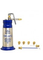
KIT DE CRIOTERAPIA CRY-AC 0.3 L
1 025,00 €
-
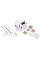
LAPARO ASPIRE VER. MEDIUM. LAPAROSCOPIC TRAINER
680,50 €
-

ADAPTADOR DE CORRIENTE ELECTRICA OMRON M2, M3, M6 IT, M7, M10 IT
19,80 €
-
FUNDA FONENDOSCOPIO CARE COLLECTION
9,95 €
-
FUNDA FONENDOSCOPIO VIRUS COLLECTION
9,95 €
-
FUNDA FONENDOSCOPIO EIGHTIES COLLECTION
9,95 €
-

GUIA PRACTICA PARA PROFESORES DE MEDICINA
72,20 € -5% 76,00 €
-

LAPARO ASPIRE VER. ESSENTIAL LAPAROSCOPIC TRAINER
638,28 €
-

FUNDA FONENDOSCOPIO ORGANOS
9,95 €


LibreriaMedica.es
 Conócenos

Estudiantes
 Estudiantes